Keywords: Open Access; Free software; Cardiac cine-MRI; Brain; Segmentation; Tagging; Cardiac deformation analysis
Overview
 his informal post presents a quick survey of what’s available for one to find when it comes to heart tagging downloads (without having to go through radiologists and all sorts of burdensome forms). It is a problem I’ve been tackling recently and I would like to share my findings to make life easier for other scientists who find themselves in the same situation.
Some Background Information
 WEEK OR SO ago I decided to share information about the availability of datasets for researchers. When faced with the problem of looking for, say, ‘”tagged MR” images heart download dicom’ (and a lot of searching around this phrase) I was surprised to find that almost nobody publicly shared or made discoverable a dataset one can use for research purposes. In order to simplify the task for others who find themselves in a similar situation, several days ago I wrote about my intentions to make a sort of summary [1, 2].
As a bit of background, our original plan was to write and implement special protocols with engineers at the MRI scanner, as part of the research effort by which special types of data are obtained for a novel approach to be tested. This is explained in a paper I’ll put up in the site pretty soon.
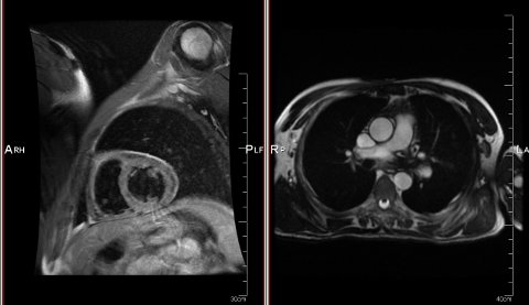
At the moment, due to particular constraints, we are working on analysis of sequences of tagged data. This obviously requires not just MR images of the heart but also sequences and these, preferably sequences that must have particular types of tags which a radiologist would use. Data can be acquired/sliced from different angles and obtained from any subject/patient at the hospital*, but for this particular approach that we test (to be covered in a separate post) this is rather irrelevant as we want it to be applicable regardless of factors like angles, subjects, and potentially their atrophies.
Resources
SourceForge hosts quite a few MRI-related software projects which are free/libre and thus of value to MRI-based research (our methods and data should be made freely accessible for independent validation). One of these projects is the cardiac MR toolbox for Matlab, which may also be fully compatible with Octave (free/libre software). “The cardiac MR (CMR) toolbox for Matlab,” explains the official Web page, “provides tools for image registration, perfusion analysis, segmentation, and image reconstruction from sparsely sampled data.” Here is the alternative project homepage. It states:
This is the Cardiac MR toolbox for Matlab project (“cmr-toolbox”)
This project was registered on SourceForge.net on Mar 29, 2010, and is described by the project team as follows:
The cardiac MR (CMR) toolbox for Matlab provides tools for image registration, perfusion analysis, segmentation, and image reconstruction from sparsely sampled data.”
For brain MRI there is the RESCUE brain MRI segmentation project. It is not yet available, but it soon will be. Its author states: “RESCUE will be made available after publication of the PhD thesis of James Withers (Uni of Edinburgh, UK). The first version of the package will initially include super-resolution, thin structure detection, and partial volume estimation components.”
Of course, one can get brain datasets from the rather famous BrainWeb (more here) and some more data resides in my site, including a sample set of images and some DICOM-formatted data of my own brain (acquired at Hope Hospital for a colleague’s experiment back in 2005).
Moving on to the heart, there is a cardiac MRI dataset which is not terribly useful because almost everything is encoded and stored in MATLAB format and may therefore require some loaders that are proprietary (Octave does not cope so well with those).
There is another project called inTag and this Web pageoffers a a look at what’s done there. Another page offers a look at how it’s used. It is “a software to calculate, display and analyze myocardial strains and intra-myocardial mechanics from cardiac MR images with a tagging pattern.”
The dependencies on proprietary software are unfortunate though. As the page puts it: “After a preliminary prototyping step in Matlab ®, InTag is now available as a plugin in OsiriX, the advanced open-source image processing software. It benefits of all its unique capabilities for navigation, visualisation of multimodality and multidimensional images, and data management.”
This requires OsiriX, which is Free/open source software, but only for Macs, which is not terribly useful then. Here is some tagging from this software package called inTag. It’s just a group of some very low-resolution images in DICOM format (small and not so useful at all).
Another dataset which I find odd (not because it’s made available just in very low resolution but because there is something odd in orientation) comes from New Zealand. Images are said to be available for download, but they are all encoded as AVI-formatted slanted videos and also compressed. The only one set with tagging data is not too helpful, either. The page states that “The Auckland MRI Research group has agreed to make freely available 256×256 JPEG images and image orientation information for download. In addition the Auckland MRI Research group has also agreed to make freely available the cines in AVI format”. What’s listed in the page as an overview is as follows:
All 119 T1-weighted jpeg images
akld-mrirg-atlas-jpegs.zip (4.33MB)
12 Long axis trueFisp avi movies
akld-mrirg-cines-truefisp-long.zip (21.76MB)
1-15 Short axis trueFisp avi movies
akld-mrirg-cines-truefisp-short-a.zip (25.32MB)
16-30 Short axis trueFisp avi movies
akld-mrirg-cines-truefisp-short-b.zip (28.61MB)
3 Aorta and 1 RVOT trueFisp avi movies
akld-mrirg-cines-truefisp-aorta-rvot.zip (7.42MB)
11 Long axis and 15 Short axis tagged avi movies
akld-mrirg-cines-tagging.zip (26.46MB)
Aorta(2) / Right(2) and Left(2) Pulmonary arteries(4) phase contrast avi
movies akld-mrirg-cines-phasecontrast.zip (10.34MB)
Those sets are not tagged except akld-mrirg-cines-tagging.zip. There is also no data from the Cincinnati Children’s Hospital Medical Center, which shows up among many search results. Search for data has proven to be quite fruitless, generally speaking.
Eventually I used data which I acquired from someone’s thesis frame by frame, only to be seen as a temporary solution until data comes from a group that keeps it for internal use. The conclusion is that the Web is generally a poor resource for such data, especially if tagging is a requirement. If someone knows of an exception, please comment below.
Some References
On this fruitless pursuit for data the following references were seen as potentially relevant (publication names mostly omitted as it’s easy to search by title).
DN Metaxas, “A Segmentation and Tracking System for 4D Cardiac Tagged MR Images,” 2008.
L Axel, “Tagged MRI-Based Studies of Cardiac Function,” Computerized Medical Imaging and Graphics, Volume 29, Issue 8, December 2005, Pages 607-616, 2003.
D Goksel, “Towards rapid screening of tagged MR images of the heart,” 2002.
NF Osman, “Angle Images for Measuring Heart Motion from Tagged MRI.”
L Axel, “An Integrated Program for 2-D and 3-D Analysis of Heart Wall…,” 2002.
Y. Chenounea, “Segmentation of cardiac cine-MR images and myocardial deformation assessment using level set methods,” 2005.
(Abstract: “In this paper, we present an original method to assess the deformations of the left ventricular myocardium on cardiac cine-MRI. First, a segmentation process, based on a level set method is directly applied on a 2D+t dataset to detect endocardial contours. Second, the successive segmented contours are matched using a procedure of global alignment, followed by a morphing process based on a level set approach. Finally, local measurements of myocardial deformations are derived from the previously determined matched contours. The validation step is realized by comparing our results to the measurements achieved on the same patients by an expert using the semi-automated HARP reference method on tagged MR images.)
____
* Some are too blurry and others paint areas with blood presence black, which help isolate the boundaries of important structures for subsequent analysis.
 fter some preparation and discussions among ourselves, we are finally ready to show SCI Fitness to the outside world. Some of the pages are still work in progress, but we are eager to make updates and provide information in the near future, so by all means subscribe to the RSS feed if interested.


 Filed under:
 ust over 15 years ago I started lifting weights. I had only just turned 14 at the time. The memories are many and it was only about 7 years ago that I started writing abut it. I can probably reference my older blog posts about health and exercise, starting with
 aving discussed the subject with half a dozen people over the past week or so, it seems clear that the threat of radiation is underplayed by companies that make business out of it (X-ray for the most part, if not nuclear energy too). The crux of the argument is that, just as people are increasingly not allowed to smoke in pubs due to people who work there (not always out of choice), in places where radiation is abundant, e.g. scanning facilities in hospitals and airports, people’s right to deny and avoid exposure to radiation should be respected. In companies where CT scanners are tested, even software developers are required to be exposed to radiation (my siblings) and sign a sort of waiver that removes liability. In more and more airports not only suitcases are subjected to X-ray treatment but humans too; they are not even given alternative options, except not work (in the former case) or not fly (in the latter case). The attitude ought to change. As someone with doctoral qualifications in the area of medical imaging, I occasionally try to raise the issue (not confront) those who are victim of this status quo (e.g. hospital workers who spend hours in particular rooms and particular airport staff standing adjacent to high-power scanners), but it is not easy to reach a solution which does not leave both sides with relative discomfort.
 am well past my
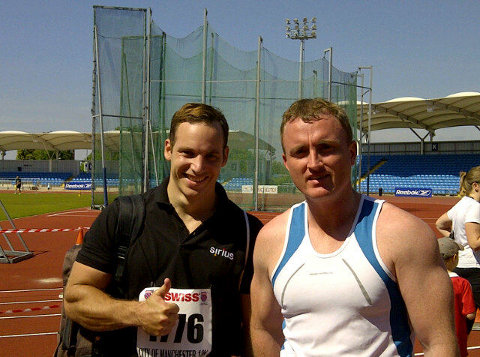
 S Independence Day/Weekend was a lot of fun. The Manchester City Stadium 10K (ten kilometers, which is about a quarter of a marathon) took place on Sunday. I was among the runners there and the following day I cycled for 9 hours because it was nice and sunny (now it rains again). Today I spent over 3 hours at the gym, making the total of time spent on exercise in the past 3 days something around 16 hours. In order to keep in decent shape but not make races just a torturous experience (I went to the gym straight afterwards for weights), I took it easy in this race and I intend to carry on running and cycling more than before, maybe building up stamina for a change. My diet too has improved somewhat. So anyway, it was a very pleasant weekend, maybe I will upload more photos later. I will probably do the same Manchester 10K race next year as it exceeded my expectations. The number of participants was a couple of thousands, meaning far smaller than the great run, which is also 10K but has a low of slow runners. The race was by mere chance talking place on one of the more pleasant days of the year, even though it was a little too hot for running (early in the day and not a cloud in the sky). Anyone who considers doing future K-Swiss Manchester 10K races probably won’t regret it.
 his informal post presents a quick survey of what’s available for one to find when it comes to heart tagging downloads (without having to go through radiologists and all sorts of burdensome forms). It is a problem I’ve been tackling recently and I would like to share my findings to make life easier for other scientists who find themselves in the same situation.
