Monday, October 25th, 2010, 8:50 am
Cardiac References
 HIS DRAFT-LEVEL OR DRAFT-QUALITY POST is not a detailed literature review but only an attempt to group together papers of interest and highlight those which are potentially relevant to in vivo studies of the heart, such as studies that distinguish between a healthy heart and a diseased one based on its motion. In this work, the goal is to study the registration and/or analysis of heart images and how one can overcome the 3 types of movement which occur at a rapid rate with less than one second per cycle, where the heart twists and shrinks (changes in volume), thus leading to ambiguity and confusion. The muscle tissue inside the heart and outside the heart is positioned in such a way that fibre sticks at a roughly 60-degree angle perpendicular to the heart, with the outside being at odds wrt to the angle that’s seen on the inside. Things change when blood is being pumped.
My initial goal revolves around 3-D heart reconstruction, tracing/tracking cardiac activity, attempting an approach of an isolated cube with occlusion to track one position of the heart amid movement (if that fails, there is another simpler shape analysis approach from around 1990).
I have been shifting focus from my background in analysis of brain MRI (and to a limited extent exploration of face images for recognition of sets and statistical analysis of these). Cardiac 3-D is a subject which may require an introduction to the heart and its dynamics in particular.
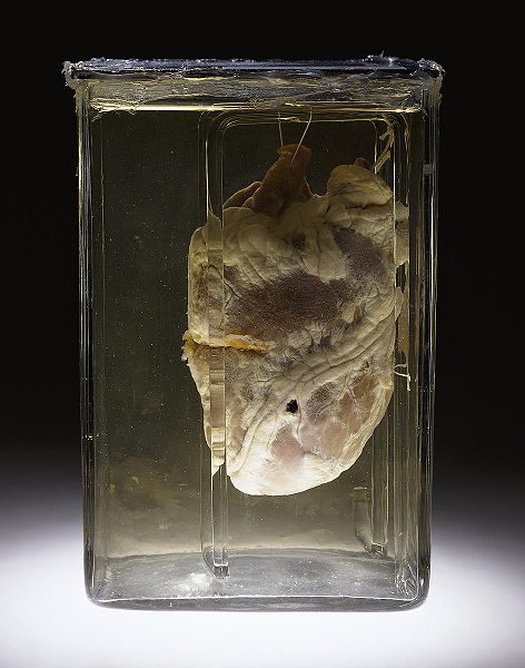
The Wikipedia article which focuses mostly on the human heart clarifies that the term cardiac (as in cardiology) means “related to the heart” and comes from the Greek kardia, which can be almost be substituted to mean “heart”. Some useful pages from the University of Minnesota are worth a look. Here is a nice interactive map with which to study the anatomy of the heart but not its operation. Cardiac anatomy can be learned from it. Lastly, the Atlas of Human Cardiac Anatomy allows viewing the heart as it pumps blood. It is animated. Here is static photo of interest.
The unfortunate thing is that I remain dependent on MATLAB, which is the only proprietary piece of software (working properly on GNU/Linux though) I remains stuck with while doing my research, partly for collaboration purposes, which is ironic when the underlying framework is all secret and from a company that funds the BSA. It’s adverse to collaboration (I was once ranked first in the world for my MATLAB contributions in MATLAB Central and was offered to write a book when approached by MathWorks, which later tried to hire me). Anyway, digressing a bit, I will be still working on DICOM datasets, just as I was doing before.
Recent papers worth considering are [1] from Isola et al., who use an NRR approach similar to the one I practiced when handling MRI images of the human brain, where motion does not exist (or barely exists under normal conditions). It is applied to CT-acquired images and not to MRI though. The abstract sums up the purpose as follows: “Cardiac computed tomography is a rapidly emerging technique for noninvasive diagnosis of cardiovascular diseases. Nevertheless, the cardiac motion continues to be a limiting factor. Electrocardiogram-gated cardiac computed tomography reconstruction methods yield excellent results, but these are limited in their temporal resolution due to the mechanical movement of the gantry, and lead to residual motion blurring artifacts. If the motion of the cardiac region of interest is determined, motion compensated gated reconstructions can be applied to reduce motion artifacts. In this paper it is shown that elastic image registration methods can be an accurate solution to determine the cardiac motion.”
Another paper [2] which has only just been published looks at three-dimensional phase-contrast MRI and results included an experiment over the cardiac phase. In an attempt to find a comprehensive review of something which better fits my background (it involved a great deal of shape and intensity models) I came across last year’s review of shape models for 3-D image segmentation [3]. In efforts to identify a cardiac literature survey or background about the heart (be it a simple article or a literature survey), I found a paper from Zhuang et al. [4] about cardiac MRI for segmentation of the heart without human intervention. It does not quite deal with the problem of motion and real-time or offline tracking of parts of the heart (4-D), though. Motion caused by respiration is studied in [5] and it is relevant to the subject of tracking and its associated perils. There are many papers about tracking in all sorts of contexts and half a year ago there was a speckle-tracking echocardiographic study into myocardial deformation from the University of Hong Kong [6]. Having looked for material on in vivo tagging, I pinned down an article from 2003 [7] on “in vivo assessment of regional myocardial work in normal canine hearts using 3D tagged MRI.”
Other authors, having studies myocardial strains over a decade earlier [7], more recently looked at what they described as a “non-invasive method for assessing regional myocardial” [8] (in 2003). Cardiac tagging was done in Johns Hopkins University last year; namely, it was myocardial tissue tagging [9] with cardiovascular magnetic resonance (CMR). More on CMR tagging (from 2008) is to be found in [10] where mouse heart is studied. A paper from Wei et al. also considered CMR tagging by experimenting on mice [11]. Methods involved “[i]n vivo myocardial function was evaluated by 3D CMR tagging in mdx mice.”
The material on CMR tagging may be of high relevance and belonging to such a literature review would be [12], which “introduce[s] a standardized method for calculation of left ventricular torsion by CMR tagging and [determining] the accuracy of torsion analysis in regions using an analytical model.” Delling et al. [13] look at CMR and explain: “We sought to assess the correlation between mitral valve characteristics and severity of mitral regurgitation (MR) in subjects with mitral valve prolapse (MVP) undergoing cardiac magnetic resonance (CMR) imaging.”
In a very recent article warning about T2-weighted CMR, a detailed explanation is given which warns that, among other things, “[i]nconsistencies also exist in the clinical literature, and we still await multicenter validation of robustness, reproducibility, and additive clinical value.” This is irrelevant to the subject at hand, but it is also exceptionally recent. This was published by Nature and the headline was somewhat alarmist: “Molecular imaging: T2-weighted CMR of the area at risk—a risky business?”
Some less relevant papers on the subject may include [14]-[15] on CMR tagging. The problem of heart tracking or tracing of parts of the heart is a complicated one and a particular group has proposed “a novel dynamic model, based on the equation of dynamics for elastic materials and on Fourier filtering.”[16]
To some groups, robotic “intervention on the beating heart” is said to be a worthy route of exploration as well [17]. This one study on pig hearts [18] — like [6] — considers speckle-tracking.
Going through some of the latest material at jcmr-online.com (which is Open Access for the most part), one can also perform paper search around “cardiac tagging”, “mri tagging” and “heart mri tagging”. This yields not so many results. There’s a highly cited paper from approximately 20 years ago titled “Cardiac Tagging with Breath-Hold Cine MRI” (Open Access). If one is looking for an article which is/contains an overview of existing strands of work, there’s work to be done, but introduction sections may often help. There is a lot about HARP, including a paper from last year [19]. It is about a method which avoid the time-consuming post-processing associated with MRI images that were tagged.
Lastly, Cardiac Imaging has this new paper from 2010, which it makes viewable as HTML, not just to subscribers to the literature [20].
It is possible that I will make some posts on this subject in the future. My other two scientific blogs are in need of a makeover as they have not been updated for years. As stated at the start, this post is informal and part of an ongoing exploration of existing work in my research area.
References:
[1] “Fully automatic nonrigid registration-based local motion estimation for motion-corrected iterative cardiac CT reconstruction.”
Author(s): Isola, Alfonso A, Grass, Michael, Niessen, Wiro J
Source: Med Phys Volume: 37 Issue: 3 Pages: 1093-109 Published: 2010
[2] Title: “In vivo hemodynamic analysis of intracranial aneurysms obtained by magnetic resonance fluid dynamics (MRFD) based on time-resolved three-dimensional phase-contrast MRI.”
Author(s): Isoda, Haruo; Ohkura, Yasuhide; Kosugi, Takashi, et al.
Source: Neuroradiology Volume: 52 Issue: 10 Pages: 921-8 Published: 2010 Oct
[3] “Statistical shape models for 3D medical image segmentation: A review”
Author(s): Heimann, T; Meinzer, HP
Source: MEDICAL IMAGE ANALYSIS Volume: 13 Issue: 4 Pages: 543-563 Published: 2009
[4] Title: “A registration-based propagation framework for automatic whole heart segmentation of cardiac MRI.”
Author(s): Zhuang, Xiahai, Rhode, Kawal S, Razavi, Reza S, et al.
Source: IEEE Trans Med Imaging Volume: 29 Issue: 9 Pages: 1612-25 Published: 2010 (EPubDate 2010 08)
[5] “Respiratory motion correction in gated cardiac SPECT using quaternion-based, rigid-body registration.”
Author(s): Parker, Jason G, Mair, Bernard A, Gilland, David R
Source: Med Phys Volume: 36 Issue: 10 Pages: 4742-54 Published: 2009
[6] Myocardial deformation in patients with Beta-thalassemia major: a speckle tracking echocardiographic study.
Author(s): Cheung, Yiu-fai; Liang, Xue-cun; Chan, Godfrey Chi-fung; Wong, Sophia Jessica; Ha, Shau-yin
Source: Echocardiography Volume: 27 Issue: 3 Pages: 253-9 Published: 2010 Mar
[7] “Distribution of myocardial strains: an MRI study.”
Author(s): Azhari, H; Weiss, J L; Shapiro, E P
Source: Adv Exp Med Biol Volume: 382 Pages: 319-28 Published: 1995
[8] “Rapid MR imaging by sensitivity profile indexing and deconvolution reconstruction (SPID).”
Author(s): Azhari, Haim; Sodickson, Daniel K; Edelman, Robert R
Source: Magn Reson Imaging Volume: 21 Issue: 6 Pages: 575-84 Published: 2003 Jul
[9] “Myocardial tissue tagging with cardiovascular magnetic resonance.”
Author(s): Shehata, Monda L; Cheng, Susan; Osman, Nael F; Bluemke, David A; Lima, Joao A C
Source: J Cardiovasc Magn Reson Volume: 11 Pages: 55 Published: 2009
[10] Title: Characterization of three-dimensional myocardial deformation in the mouse heart: an MR tagging study.
Author(s): Zhong, Jia, Liu, Wei, Yu, Xin
Source: J Magn Reson Imaging Volume: 27 Issue: 6 Pages: 1263-70 Published: 2008
[11] Title: “Early manifestation of alteration in cardiac function in dystrophin deficient mdx mouse using 3D CMR tagging.”
Author(s): Li, Wei, Liu, Wei, Zhong, Jia, et al.
Source: J Cardiovasc Magn Reson Volume: 11 Pages: 40 Published: 2009 22
[12] “Regional assessment of left ventricular torsion by CMR tagging.”
Author(s): Russel, Iris K; Gotte, Marco J; Kuijer, Joost P; Marcus, J Tim
Source: J Cardiovasc Magn Reson Volume: 10 Pages: 26 Published: 2008
[13] “Title: CMR Predictors of Mitral Regurgitation in Mitral Valve Prolapse.”
Author(s): Delling, Francesca N, Kang, Lih Lisa, Yeon, Susan B, et al.
Source: JACC Cardiovasc Imaging Volume: 3 Issue: 10 Pages: 1037-45 Published: 2010
[14] “Altered myocardial motion pattern in Fabry patients assessed with CMR-tagging.”
Author(s): Rutz, Andrea K, Juli, Christoph F, Ryf, Salome, et al.
Source: J Cardiovasc Magn Reson Volume: 9 Issue: 6 Pages: 891-8 Published: 2007
[15] Title: “Septal ablation in hypertrophic obstructive cardiomyopathy improves systolic myocardial function in the lateral (free) wall: a follow-up study using CMR tissue tagging and 3D strain analysis.”
Author(s): van Dockum, Willem G, Kuijer, Joost P A, Gotte, Marco J W, et al.
Source: Eur Heart J Volume: 27 Issue: 23 Pages: 2833-9 Published: 2006 (EPubDate 2006 10)
[16] “A dynamic elastic model for segmentation and tracking of the heart in MR image sequences.”
Author(s): Schaerer, Joel; Casta, Christopher; Pousin, Jerome; Clarysse, Patrick
Source: Med Image Anal Volume: 14 Issue: 6 Pages: 738-49 Published: 2010 Dec
[17] “Collaborative tracking for MRI-guided robotic intervention on the beating heart.”
Author(s): Zhou, Y; Yeniaras, E; Tsiamyrtzis, P; Tsekos, N; Pavlidis, I
Source: Med Image Comput Comput Assist Interv Volume: 13 Issue: Pt 3 Pages: 351-8 Published: 2010
[18] “Three-dimensional speckle-tracking imaging for left ventricular rotation measurement: an in vitro validation study.”
Author(s): Zhou, Zhiwen; Ashraf, Muhammad; Hu, Dayi; Dai, Xiaonan; Xu, Yawei; Kenny, Bill; Cameron, Berkley; Nguyen, Thuan; Xiong, Li; Sahn, David J
Source: J Ultrasound Med Volume: 29 Issue: 6 Pages: 903-9 Published: 2010 Jun
[19] “Transmural myocardial strain in mouse: quantification of high-resolution MR tagging using harmonic phase (HARP) analysis.”
Author(s): Zhong, Jia, Liu, Wei, Yu, Xin
Source: Magn Reson Med Volume: 61 Issue: 6 Pages: 1368-73 Published: 2009
[20] “Quantification in cardiac MRI: advances in image acquisition and processing”
Anil K. Attili, Andreas Schuster, Eike Nagel, Johan H. C. Reiber and Rob J. van der Geest
The International Journal of Cardiovascular Imaging (formerly Cardiac Imaging)
Volume 26, Supplement 1, 27-40


 Filed under:

